您好!歡迎訪問洛陽富道生物科技有限公司官方網(wǎng)站!


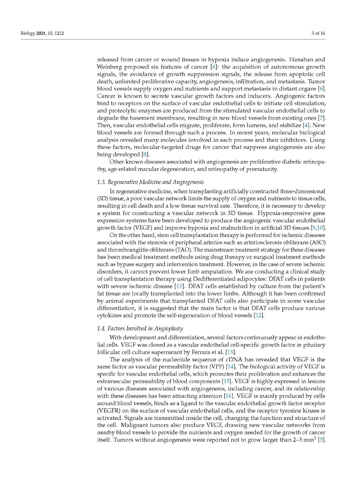
The control of angiogenesis is essential in disease treatment or regenerative medicine. We conducted a clinical study of dedifferentiated fat (DFAT) cells, a kind of mesenchymal stem cells, by applying cell transplantation therapy to induce angiogenesis in patients with severe ischemic disease. This study aimed to analyze the effect of molecules that regulate angiogenesis in vitro and clarify their molecular mechanisms for therapeutic purposes. Normal human umbilical venous endothelial cells (HUVECs) were cultured in the presence of vascular endothelial growth factor (VEGF). Recombinant human angiopoietin-1-producing cells, conditioned media, mouse DFAT cells, and antioxidant polyphenols were added to this system at various concentrations. After 11 days, the cultures were immunostained with CD31 (PECAM-1), and microscopic images were subjected to analysis (area, length, joint, and path) by using software to quantitatively analyze blood vessel formation. The expression of angiogenic markers and COX pathway genes were analyzed by RT-PCR. As a result, the dose-dependent angiogenesis-promoting effect of rAng-1-producing cells, conditioned medium, or commercially available recombinant Ang-1 were observed. DFAT cells also promoted angiogenesis, whereas polyphenols inhibited angiogenesis in a dose-dependent manner.
血管生成的控制在疾病治療或再生醫(yī)學中是必不可少的。我們通過應用細胞移植療法誘導嚴重缺血性疾病患者的血管生成,對去分化脂肪 (DFAT) 細胞(一種間充質干細胞)進行了臨床研究。本研究旨在分析體外調(diào)節(jié)血管生成的分子的作用,并闡明其用于治療目的的分子機制。在血管內(nèi)皮生長因子 (VEGF) 存在下培養(yǎng)正常人臍靜脈內(nèi)皮細胞 (HUVEC)。將產(chǎn)生不同濃度的重組人血管生成素 1 細胞、條件培養(yǎng)基、小鼠 DFAT 細胞和抗氧化多酚添加到該系統(tǒng)中。11 天后,用 CD31 (PECAM-1) 對培養(yǎng)物進行免疫染色,通過軟件對顯微圖像進行分析(面積、長度、關節(jié)、路徑),對血管形成進行定量分析。通過RT-PCR分析血管生成標志物和COX通路基因的表達。結果,觀察到了產(chǎn)生 rAng-1 的細胞、條件培養(yǎng)基或市售重組 Ang-1 的劑量依賴性血管生成促進作用。DFAT 細胞還促進血管生成,而多酚以劑量依賴性方式抑制血管生成。或可商購的重組Ang-1被觀察到。DFAT 細胞還促進血管生成,而多酚以劑量依賴性方式抑制血管生成。或可商購的重組Ang-1被觀察到。DFAT 細胞還促進血管生成,而多酚以劑量依賴性方式抑制血管生成。
2.1. Materials
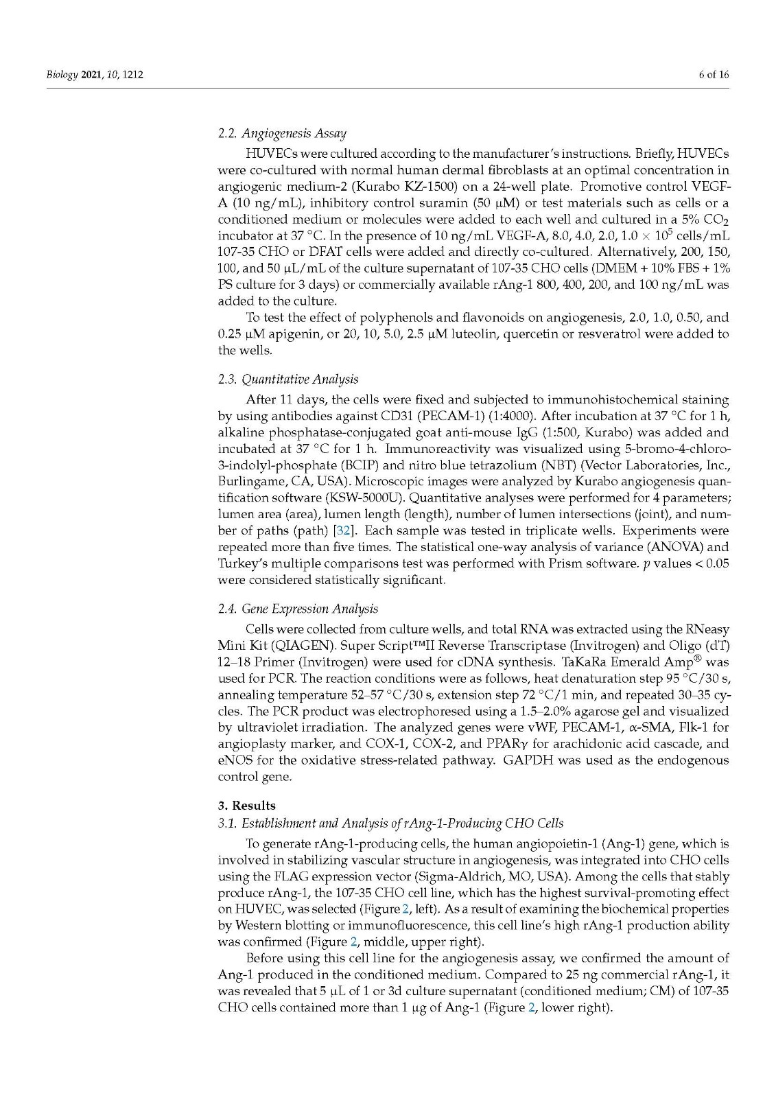
Normal human umbilical vein endothelial cells (HUVECs) were purchased as an angiogenesis kit (Kurabo, Osaka, Japan; KZ-1000). Human rAng-1-producing 107-35 CHO cells were made by NN. DFAT-D1 cells were established from mature adipocytes of adult ddY mice [31].
Polyphenols apigenin and luteolin (Fujifilm Wako Chemicals) were dissolved in methanol, resveratrol, and quercetin (Fujifilm Wako Chemicals) dissolved in ethanol. Recombinant Ang-1 protein was purchased from R&D Systems. CD31 antibody (tube formation indicator), VEGF-A (positive control), and suramin (negative control) were purchased from Kurabo.
2.2. Angiogenesis Assay
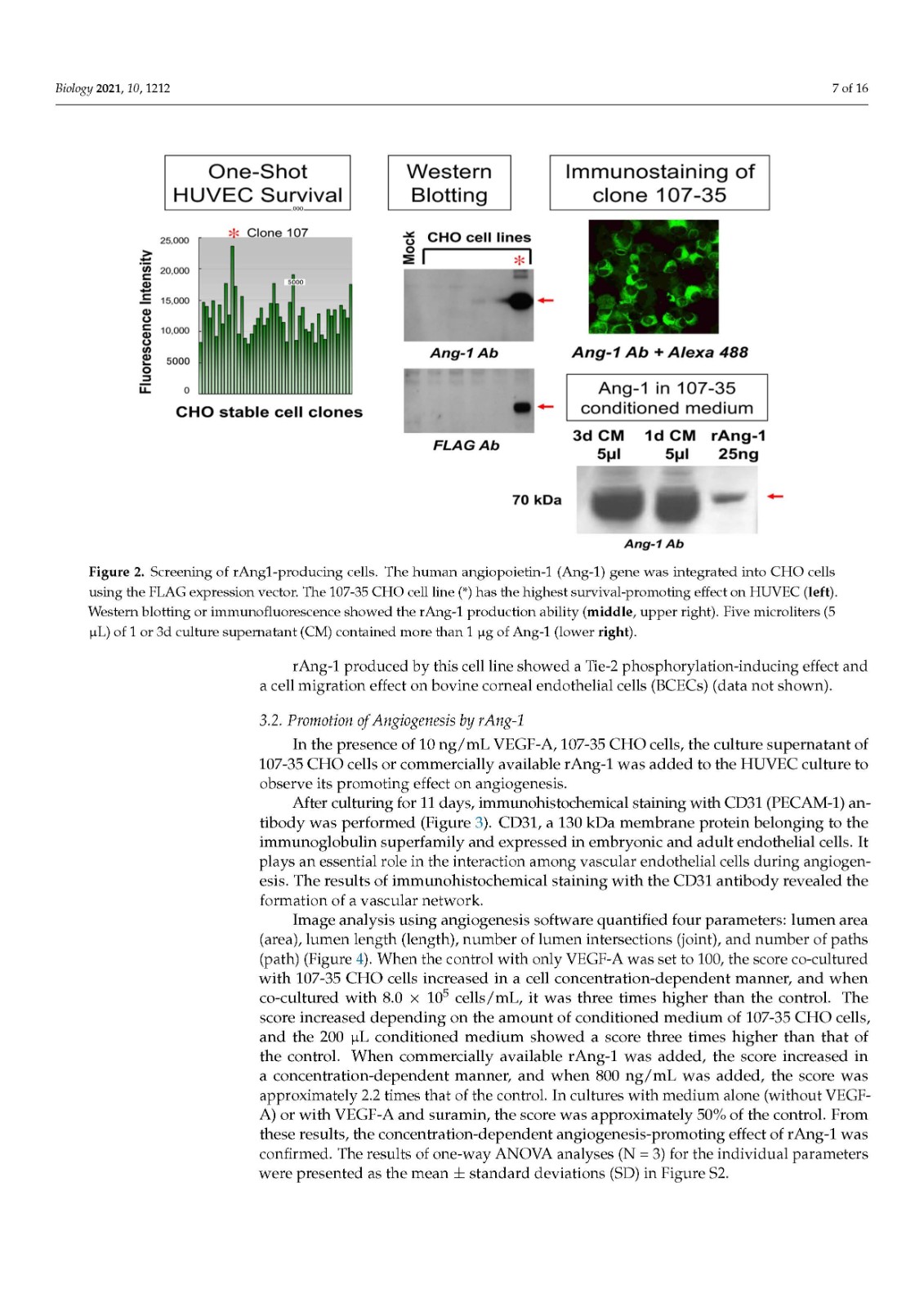
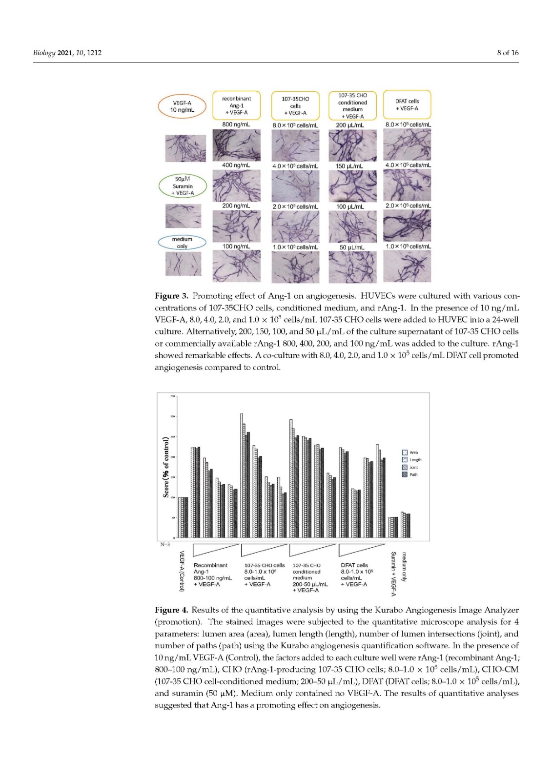
HUVECs were cultured according to the manufacturer’s instructions. Briefly, HUVECs were co-cultured with normal human dermal fibroblasts at an optimal concentration in angiogenic medium-2 (Kurabo KZ-1500) on a 24-well plate. Promotive control VEGF-A (10 ng/mL), inhibitory control suramin (50 μM) or test materials such as cells or a conditioned medium or molecules were added to each well and cultured in a 5% CO2 incubator at 37 °C. In the presence of 10 ng/mL VEGF-A, 8.0, 4.0, 2.0, 1.0 × 10⁵ cells/mL 107-35 CHO or DFAT cells were added and directly co-cultured. Alternatively, 200, 150, 100, and 50 μL/mL of the culture supernatant of 107-35 CHO cells (DMEM + 10% FBS + 1% PS culture for 3 days) or commercially available rAng-1 800, 400, 200, and 100 ng/mL was added to the culture.
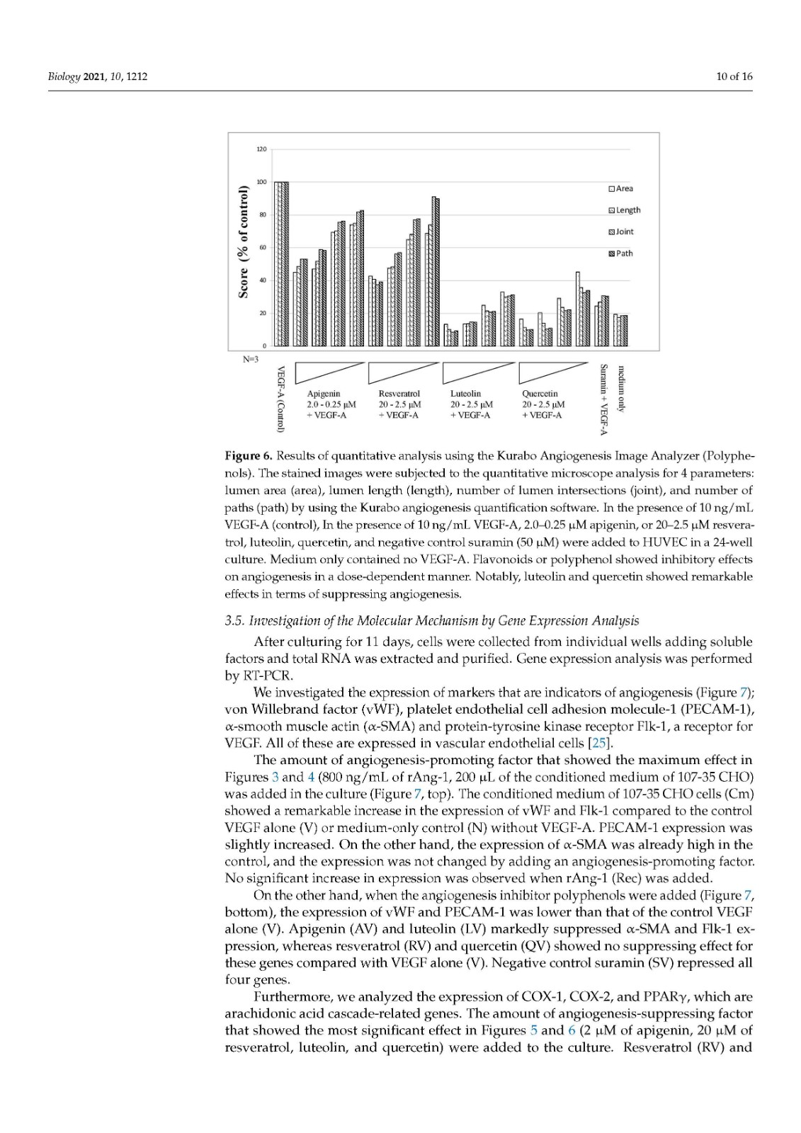
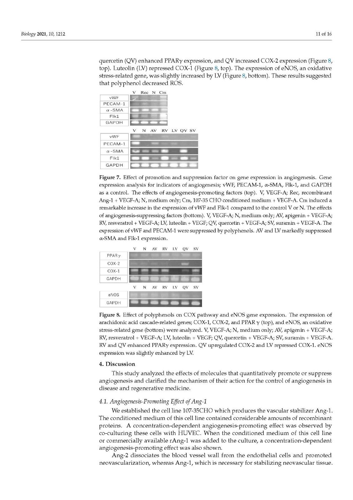
To test the effect of polyphenols and flavonoids on angiogenesis, 2.0, 1.0, 0.50, and 0.25 μM apigenin, or 20, 10, 5.0, 2.5 μM luteolin, quercetin or resveratrol were added to the wells.
正常人臍靜脈內(nèi)皮細胞(HUVEC)作為血管生成試劑盒(Kurabo,Osaka,Japan;KZ-1000)購買。產(chǎn)生人 rAng-1 的 107-35 CHO 細胞由 NN 制備。DFAT-D1 細胞是從成年 ddY 小鼠的成熟脂肪細胞建立的。
多酚芹菜素和木犀草素(Fujifilm Wako Chemicals)溶解在甲醇中,白藜蘆醇和槲皮素(Fujifilm Wako Chemicals)溶解在乙醇中。重組 Ang-1 蛋白購自 R&D Systems。CD31 抗體(管形成指示劑)、VEGF-A(陽性對照)和蘇拉明(陰性對照)購自 Kurabo。
2.2. 血管生成分析
根據(jù)制造商的說明培養(yǎng) HUVEC。簡而言之,將 HUVEC 與正常人真皮成纖維細胞以最佳濃度共培養(yǎng)在 24 孔板上的血管生成培養(yǎng)基 2(Kurabo KZ-1500)中。將促進對照 VEGF-A (10 ng/mL)、抑制對照蘇拉明 (50 μM) 或測試材料(如細胞或條件培養(yǎng)基或分子)添加到每個孔中,并在 5% CO 2培養(yǎng)箱中于 37°C 下培養(yǎng)。在 10 ng/mL VEGF-A 存在下,添加 8.0、4.0、2.0、1.0 × 10 5細胞/mL 107-35 CHO 或 DFAT 細胞并直接共培養(yǎng)。或者,200、150、100 和 50 μL/mL 的 107-35 CHO 細胞培養(yǎng)上清液(DMEM + 10% FBS + 1% PS 培養(yǎng) 3 天)或市售 rAng-1 800、400、200,并向培養(yǎng)物中加入 100 ng/mL。為了測試多酚和類黃酮對血管生成的影響,將 2.0、1.0、0.50 和 0.25 μM 芹菜素,或 20、10、5.0、2.5 μM 木犀草素、槲皮素或白藜蘆醇添加到孔中。
瓶")
T75細胞培養(yǎng)瓶
This study aimed to quantitatively analyze the effects of molecules that promote or suppress angiogenesis and clarify the mechanism for its regulation. Based on the results of numerical image analyses, the effects of Ang-1 and polyphenols on vascular tissue formation were examined. As a result, we could clarify the concentration-dependent angiogenesis-promoting effect of Ang-1 and the concentration-dependent angiogenesis-suppressing effect of four types of polyphenols. On the other hand, a co-culture with DFAT cells also showed an angiogenesis-promoting effect. We would like to apply the control of angiogenesis by the factors clarified in this study and apply it in regenerative vascular medicine.
本研究旨在定量分析促進或抑制血管生成的分子的作用并闡明其調(diào)控機制。基于數(shù)值圖像分析的結果,研究了 Ang-1 和多酚對血管組織形成的影響。結果,我們可以闡明Ang-1的濃度依賴性血管生成促進作用和四種多酚的濃度依賴性血管生成抑制作用。另一方面,與 DFAT 細胞的共培養(yǎng)也顯示出促進血管生成的作用。我們希望通過本研究闡明的因素控制血管生成,并將其應用于再生血管醫(yī)學。
HUVEC,血管生成,Ang-1,血管內(nèi)皮生長因子,類黃酮,多酚HUVECs,angiogenesis,Ang-1,VEGF,flavonoid,polypheno,DFAT
來源:MDPI https://www.mdpi.com/2079-7737/10/11/1212/htm
上一篇: 培養(yǎng)基瓶可以用來盛裝哪些溶液

統(tǒng)一服務熱線
400-160-1996
聯(lián)系人:劉英
聯(lián)系電話:13373342369
地址:河南省洛陽市洛龍區(qū)宇文愷街25號3幢101