您好!歡迎訪問洛陽富道生物科技有限公司官方網(wǎng)站!


Colorectal cancer is the third most common type of cancer in the world. Immune cells and normal supporting cells (MSCs) within a tumour affect patient survival and change how well treatments work. This research aimed to develop a more relevant 3D cancer model that combines MSCs and immune cells with cancer cells to test the effects of multiple cell types on tumour growth. We successfully developed a 3D model that shows that MSCs and immune cells can change the cancer-supporting environment around the tumour cells. We show that combining MSCs and immune cells with cancer cells can increase the level of immune-suppressing molecules they release and change immunotherapeutic drug targets on the cancer cells, similar to changes seen in human tumours. Using this 3D model for research may be better for testing new drugs than traditional 2D methods and could enable the identification of new drug targets.
結(jié)直腸癌是世界上第三大最常見的癌癥類型。腫瘤內(nèi)的免疫細胞和正常支持細胞 (MSC) 會影響患者的生存并改變治療效果。這項研究旨在開發(fā)一種更相關(guān)的 3D 癌癥模型,將 MSC 和免疫細胞與癌細胞相結(jié)合,以測試多種細胞類型對腫瘤生長的影響。我們成功開發(fā)了一個 3D 模型,顯示 MSCs 和免疫細胞可以改變腫瘤細胞周圍的癌癥支持環(huán)境。我們表明,將 MSC 和免疫細胞與癌細胞結(jié)合可以增加它們釋放的免疫抑制分子的水平,并改變癌細胞上的免疫治療藥物靶點,類似于人類腫瘤中所見的變化。
2.1. CT26 Culture and Generation of Tumour-Conditioned Medium
Mouse colon adenocarcinoma cells, CT26, derived from Balb/c mice, were purchased from the European Collection of Authenticated Cell Cultures (ECACC) and grown in CT26 media, DMEM (Gibco, ThermoFisher Scientific, Waltham, MA, USA), supplemented with 10% foetal bovine serum (FBS; Sigma-Aldrich, Wicklow, Ireland) and 1% penicillin/streptomycin (ThermoFisher Scientific, Waltham, MA, USA). Tumour-cell secretome (TCS) was generated by plating 1 × 10⁶ CT26 cells in a volume of 25 mL medium for 72 h. Then, the TCS was collected, spun at 1000 RCF for 5 min and stored at ?80 °C.
2.2. Murine MSC Isolation and Culture
Balb/c mice were purchased from Envigo Laboratories (Oxon, UK), housed and maintained following the conditions approved by the Animal Care Research Ethics Committee of the National University of Ireland, Galway (NUIG), under individual and project authorisation licenses from the Health Products Regulatory Authority (HPRA) of Ireland, in a f ushed from the bones. Cells were filtered and plated at a density of 1 × 10⁶ cells per T175 flask (Sarstedt, Wexford, Ireland) at 37 °C in normoxia (21% O2) in MEM (ThermoFisher Scientific, Waltham, MA, USA) supplemented with 10% FBS and 1% penicillin/streptomycin. Non-adherent cells were removed 24 h later through a medium change. This process was repeated until cells reached confluency. MSCs were characterised according to the criteria set out by the International Society for Cellular Therapy (ISCT) [31].
2.3. Human MSC Isolation and Culture
Human MSCs were isolated from the bone marrow from healthy volunteers at Galway University Hospital under an ethically approved protocol (NUIG Research Ethics Committee, Ref: 8 May 2014). Written consent was obtained from the volunteers. Briefly, bone marrow cell suspensions were layered onto a Ficoll density gradient, and the nucleated cell fraction was collected, washed and re-suspended in hMSC culture medium. After 24 h of culture, non-adherent cells were removed, fresh medium was added and individual colonies of fibroblast-like cells expanded. hMSCs were grown in α-MEM supplemented with 10% FBS, 1% penicillin/streptomycin and fibroblast growth factor 2 (FGF2, 1 ng/mL; Peprotech, London, England). MSCs were characterised according to the criteria set out by the International Society for Cellular Therapy (ISCT) [31].
2.4. Human Cell Line Culture
Human CRC cell lines HCT116, HT29, SW480 and the human monocyte cell line THP1 were purchased from American Type Culture Collection (ATCC). Cells were grown in human cell line media, DMEM medium supplemented with 10% FBS, 1% l-glutamine and 1% penicillin/streptomycin. All cells were confirmed mycoplasma-negative (MycoAlert, Lonza, Basel, Switzerland), expanded, frozen and used within 15 passages of testing for all subsequent experiments.
2.1. CT26 培養(yǎng)和產(chǎn)生腫瘤條件培養(yǎng)基
源自 Balb/c 小鼠的小鼠結(jié)腸腺癌細胞 CT26 購自歐洲認證細胞培養(yǎng)物保藏中心 (ECACC),并在 CT26 培養(yǎng)基、DMEM(Gibco,ThermoFisher Scientific,Waltham,MA,USA)中生長,并輔以 10 % 胎牛血清(FBS;Sigma-Aldrich,Wicklow,愛爾蘭)和 1% 青霉素/鏈霉素(ThermoFisher Scientific,Waltham,MA,美國)。腫瘤細胞分泌組 (TCS) 是通過將 1 × 10 6 CT26 細胞接種在 25 mL 培養(yǎng)基中 72 小時而產(chǎn)生的。然后,收集 TCS,以 1000 RCF 旋轉(zhuǎn) 5 分鐘并儲存在 -80 °C。
2.2. 小鼠 MSC 分離和培養(yǎng)
Balb/c 小鼠購自 Envigo Laboratories (Oxon, UK),按照愛爾蘭國立大學戈爾韋 (NUIG) 動物護理研究倫理委員會批準的條件飼養(yǎng)和維持,并獲得衛(wèi)生部門的個人和項目授權(quán)許可。愛爾蘭產(chǎn)品監(jiān)管局 (HPRA),在完全認可的動物收容設(shè)施中。對于鼠 MSC (mMSC) 分離,Balb/c 小鼠被 CO 2安樂死。股骨和脛骨被移除并清除結(jié)締組織,并從骨骼中沖洗骨髓細胞。過濾細胞,涂布在1×10的密度6在常氧(37℃21%氧氣每T175燒瓶(Sarstedt的,韋克斯福德,愛爾蘭)細胞2) 在 MEM (ThermoFisher Scientific, Waltham, MA, USA) 中添加 10% FBS 和 1% 青霉素/鏈霉素。24 小時后通過更換培養(yǎng)基去除非貼壁細胞。重復(fù)該過程直到細胞達到匯合。根據(jù)國際細胞治療學會 (ISCT) [ 31 ]制定的標準對 MSC 進行表征。
2.3. 人類 MSC 分離和培養(yǎng)
根據(jù)倫理批準的協(xié)議(NUIG 研究倫理委員會,參考:2014 年 5 月 8 日),從戈爾韋大學醫(yī)院健康志愿者的骨髓中分離出人類 MSC。獲得了志愿者的書面同意。簡而言之,將骨髓細胞懸浮液分層到 Ficoll 密度梯度上,收集、洗滌并重新懸浮在 hMSC 培養(yǎng)基中的有核細胞部分。培養(yǎng) 24 小時后,去除非貼壁細胞,加入新鮮培養(yǎng)基并擴增成纖維細胞樣細胞的單個集落。hMSC 在 α-MEM 中生長,補充有 10% FBS、1% 青霉素/鏈霉素和成纖維細胞生長因子 2(FGF2,1 ng/mL;Peprotech,London,England)。根據(jù)國際細胞治療學會 (ISCT) [ 31]制定的標準對 MSC 進行表征]。
2.4. 人類細胞系培養(yǎng)
人 CRC 細胞系 HCT116、HT29、SW480 和人單核細胞系 THP1 購自美國典型培養(yǎng)物保藏中心 (ATCC)。細胞在人細胞系培養(yǎng)基、補充有 10% FBS、1% L-谷氨酰胺和 1% 青霉素/鏈霉素的 DMEM 培養(yǎng)基中生長。所有細胞都被確認為支原體陰性(MycoAlert,Lonza,Basel,Switzerland),在所有后續(xù)實驗的 15 代測試中進行擴增、冷凍和使用。
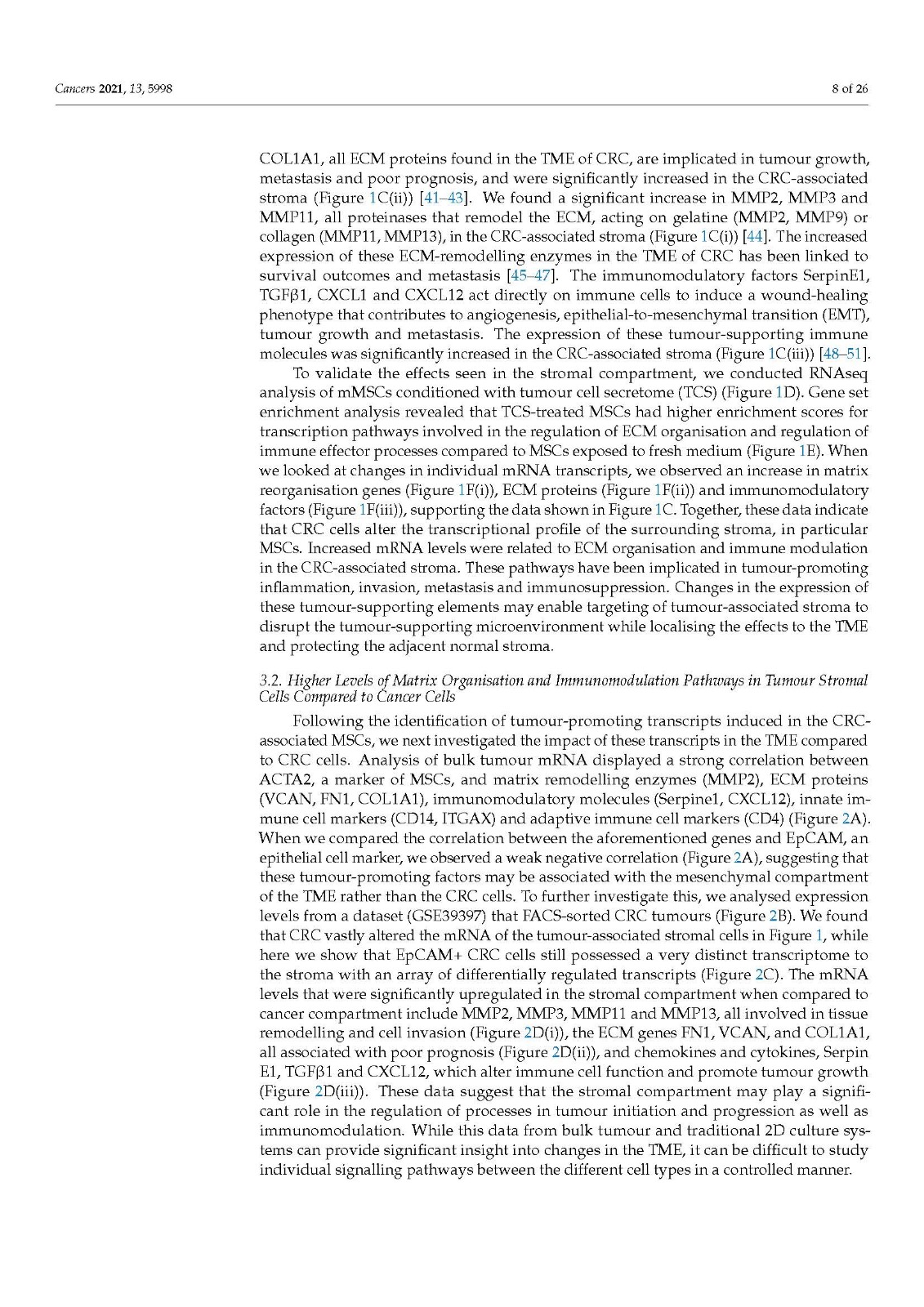
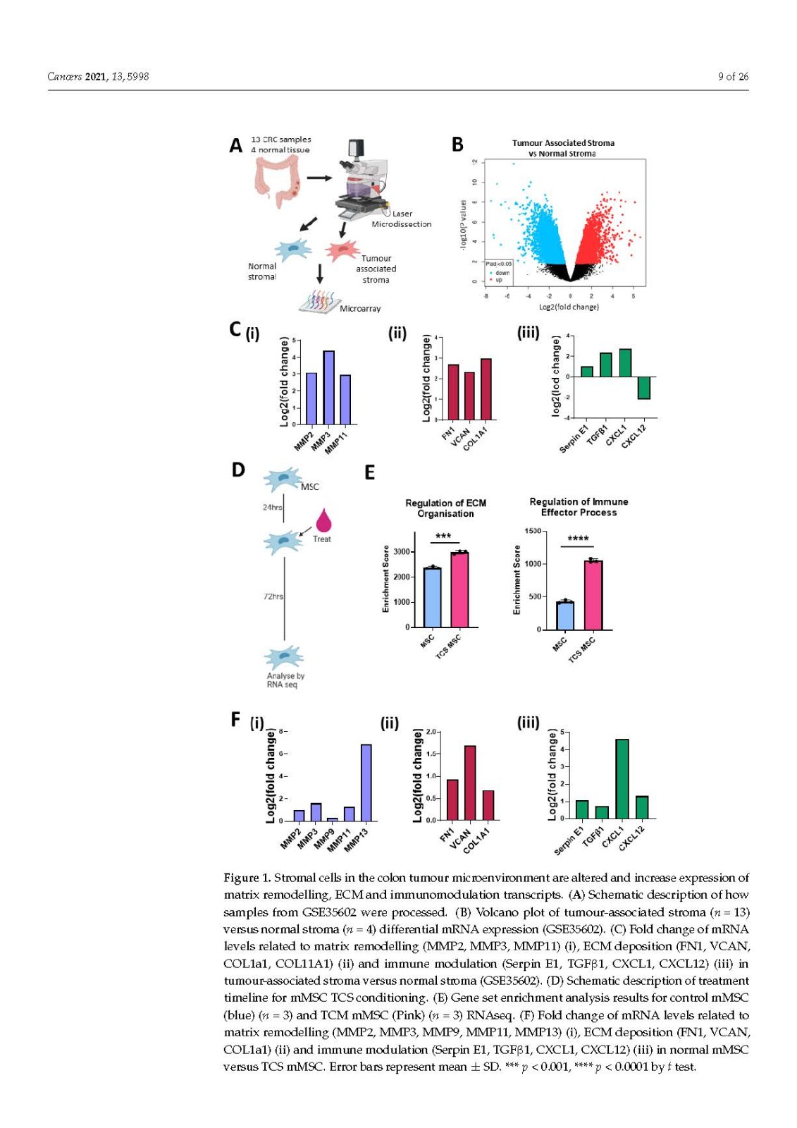
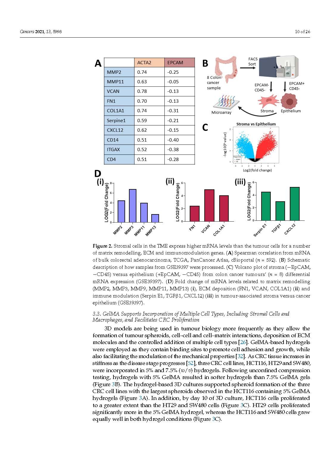
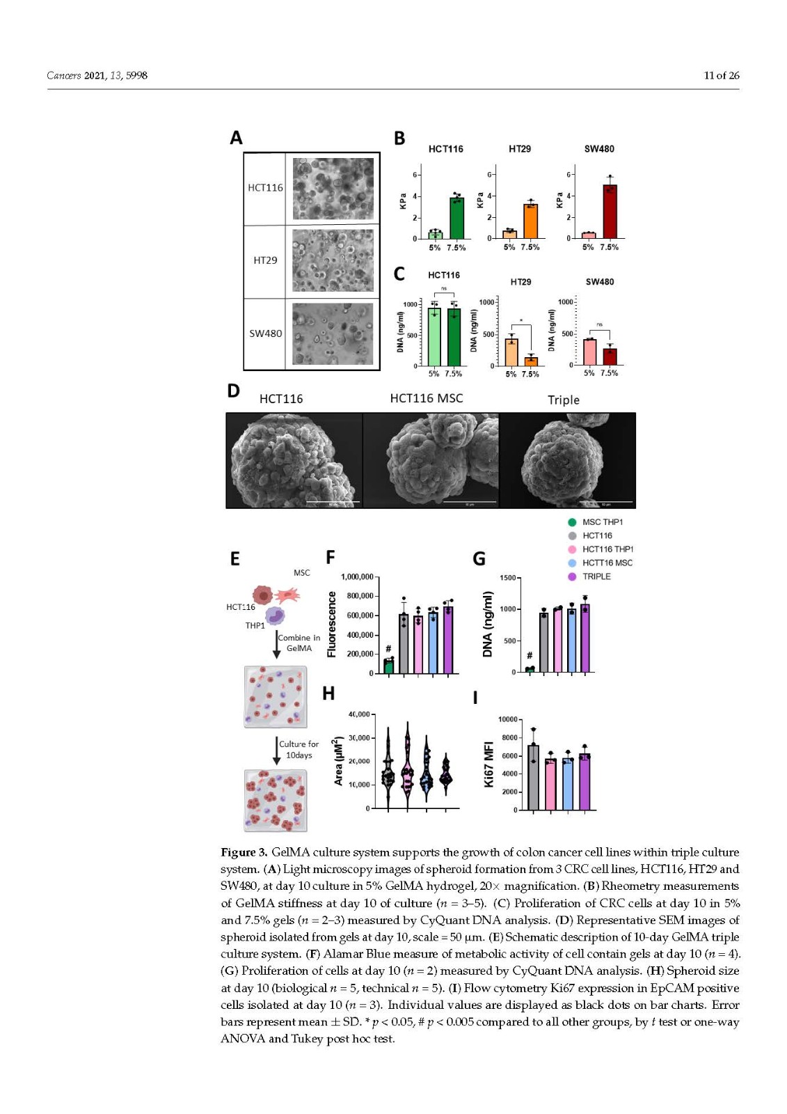
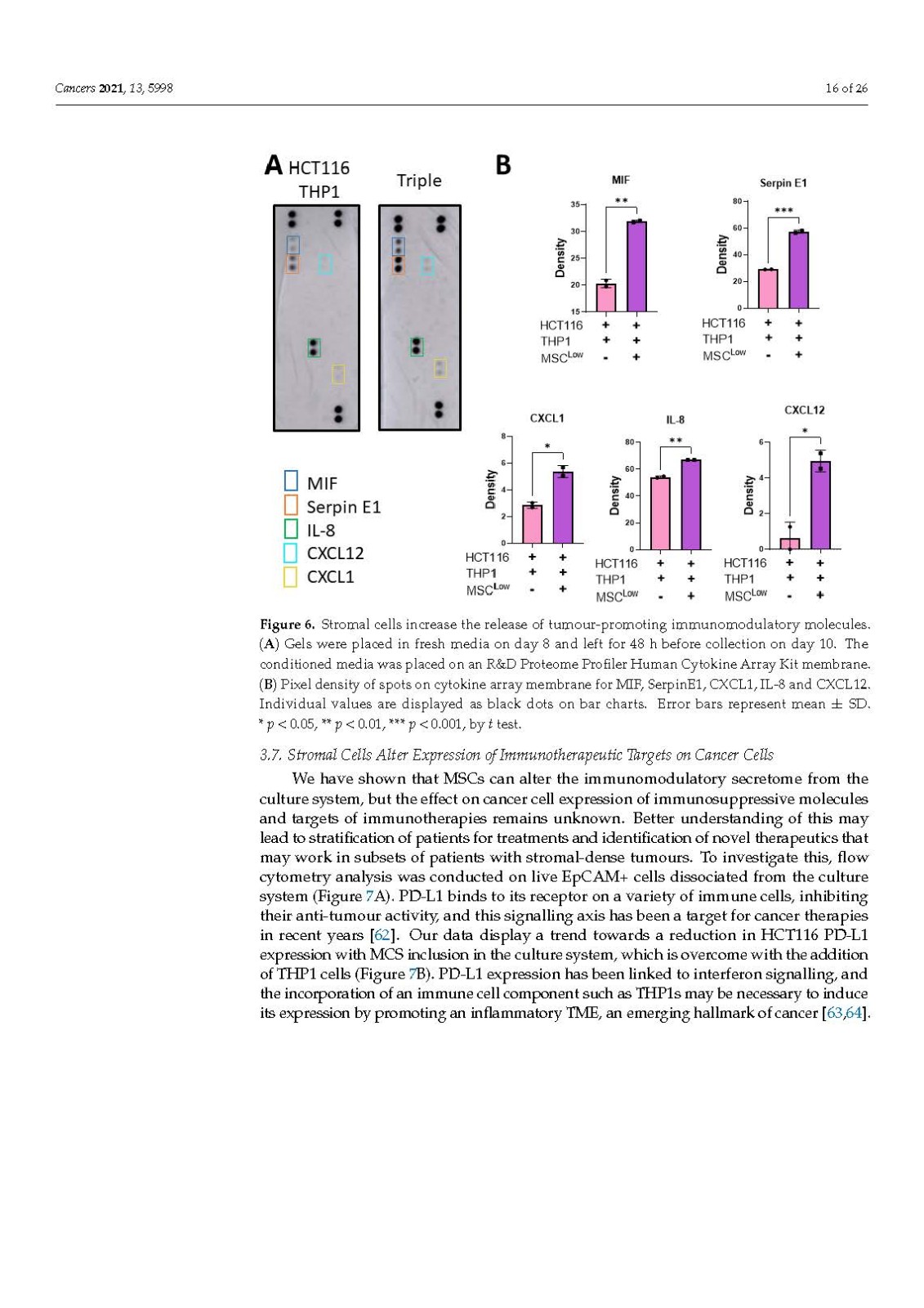
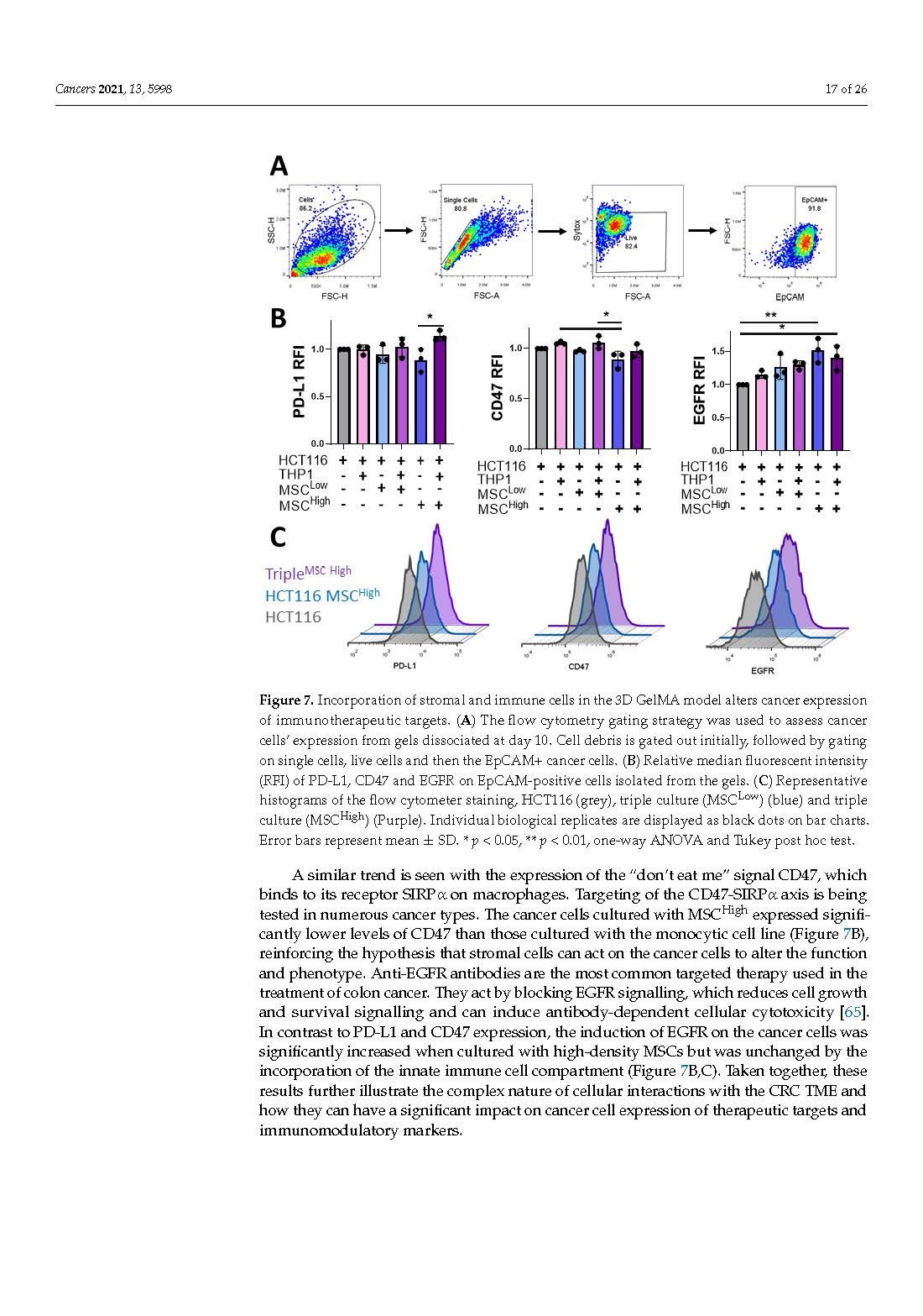
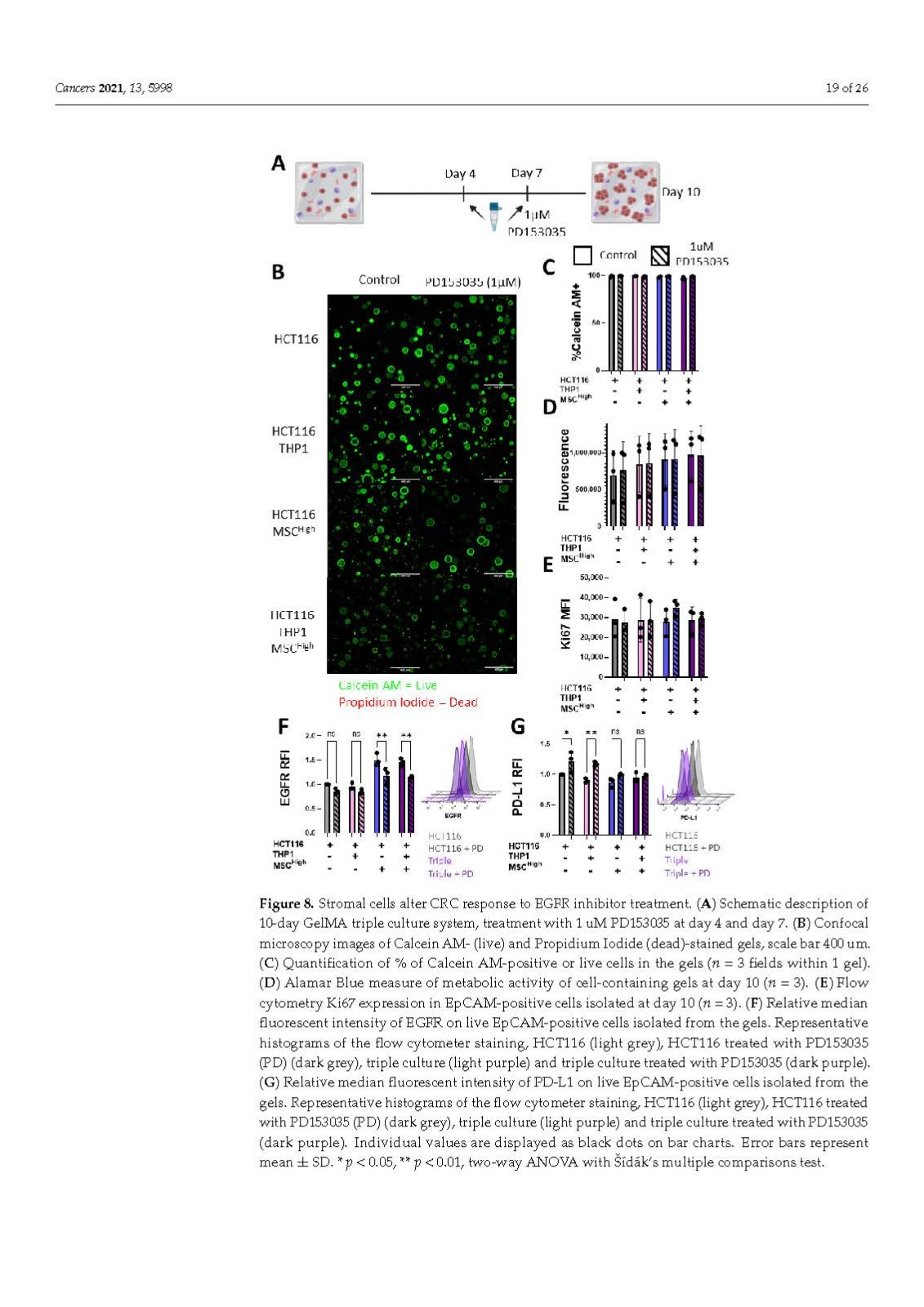
The stromal compartment of the CRC TME contributes to ECM formation, ECM remodelling, immune suppression and the induction and maintenance of an inflammatory microenvironment. These factors can alter tumour growth, metastasis, response to treatments and patient survival rates. Having accessible, reproducible models that allow the controlled modulation of each cell type is vital for the identification and testing of novel therapeutic targets and for gaining a better understanding of cellular interactions in CRC, which remains the third leading cause of cancer-related deaths worldwide. Here, we have developed a 3D triple culture model of CRC combining cancer cells, MSCs and monocytes in a tuneable hydrogel that can be analysed using a variety of techniques to assess cellular phenotype, transcriptional profiles, surface and secreted proteins. We have shown that this multicellular GelMA model has huge potential for screening targeted immunotherapeutics for CRC, due to its ease of use, reproducibility and cost effectiveness.
CRC TME 的基質(zhì)區(qū)室有助于 ECM 形成、ECM 重塑、免疫抑制以及炎癥微環(huán)境的誘導和維持。這些因素可以改變腫瘤的生長、轉(zhuǎn)移、對治療的反應(yīng)和患者的存活率。擁有允許對每種細胞類型進行受控調(diào)節(jié)的可訪問、可重復(fù)的模型對于識別和測試新的治療靶點以及更好地了解 CRC 中的細胞相互作用至關(guān)重要,CRC 仍然是全球癌癥相關(guān)死亡的第三大原因。在這里,我們開發(fā)了 CRC 的 3D 三重培養(yǎng)模型,在可調(diào)水凝膠中結(jié)合了癌細胞、MSCs 和單核細胞,可以使用各種技術(shù)進行分析,以評估細胞表型、轉(zhuǎn)錄譜、表面和分泌蛋白。
關(guān)鍵詞:colorectal cancer,tumour microenvironment,hydrogels,mesenchymal cells,extracellular matrix,3D model,immune, inflammation, stroma,結(jié)直腸癌,腫瘤微環(huán)境,水凝膠,間充質(zhì)細胞,細胞外基質(zhì),3D模型,免疫,炎癥,基質(zhì)
來源:MDPI https://www.mdpi.com/2072-6694/13/23/5998/htm


統(tǒng)一服務(wù)熱線
400-160-1996
聯(lián)系人:劉英
聯(lián)系電話:13373342369
地址:河南省洛陽市洛龍區(qū)宇文愷街25號3幢101

瓶")